Sunday, May 3, 2015

The Dissection of the Small and Large Intestine

April 15th

Focus of Procedure: Remove the small intestine and study its length and function.

Materials:
-Scalpel
-Tweezers
-Measuring Tap
-Paper Towel(s)

Procedure:
1) If you have not already done so, remove the greater and lesser momentum, which is the lining over the intestines. Removing this will provide you with a better view of the organs in the peritoneal cavity (digestive cavity). Include a picture of the greater and lesser momentum.
2) Locate the small intestine in the peritoneal cavity and identify the mesentery. The mesentery is a fold of membranous tissue that arises from the posterior wall of the peritoneal chest to the intestinal tract. Take a picture of the mesentery still attached to the intestines.
3) Once you have taken a picture, use your scalpel to cut off the mesentery from the small intestine, then throw the mesentery away.
4) Now, place the small intestine neatly back into the peritoneal cavity and take a picture of it still attached to the posterior wall of the peritoneal chest.
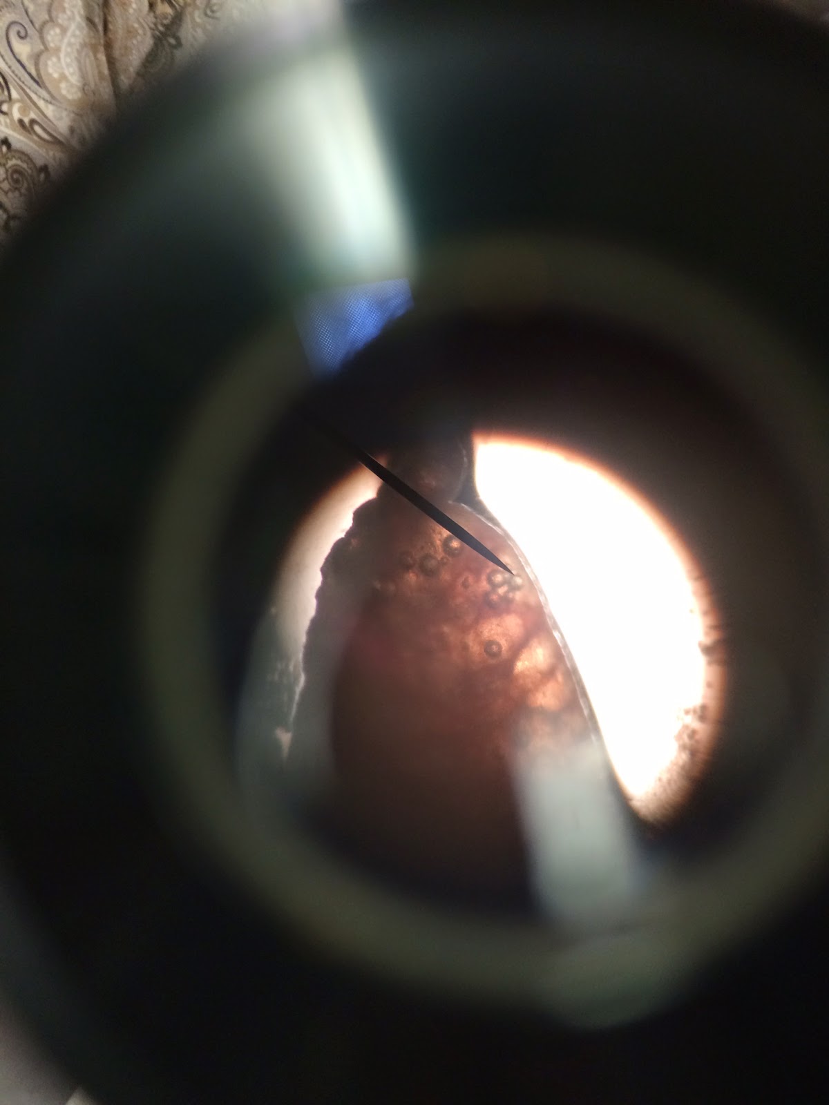
5) Please note that the small intestine has three parts: the duodenum, the jejunum and, the ilium. Re-identify the pyloric sphincter, which is where you disconnected the stomach from the small intestine. Take a picture of the duodenum (first part of the small intestine) and include a picture of the pyloric sphincter. The duodenum is the smallest part of the small intestine and begins right at the pyloric sphincter.
6) Find the end of the small intestine that connects to the large intestine. Using your scalpel, disconnect the small intestine from the large intestine, directly at the the ileocecal sphincter. The ileocecal sphincter connects the small intestine to the large intestine. Take a picture. Located in the same area, is the cecum, the first section of the large intestine that passes the digested liquid from the ileum to the colon. Take a picture.
7) Remove the small intestine from the peritoneal cavity and lay it out on a paper towel (you may need several paper towels). Completely stretch out the small intestine from end to end. Record its length in either centimeters, inches or feet. Use your measuring tape to determine the length of the small intestine and then take a picture of the small intestine completely stretched on the paper towel(s).
8) Now, locate the large intestine and take a picture. Do not remove the large intestine from the peritoneal cavity, you will only need to pictures the parts of the large intestine. The large intestine, otherwise known as the colon, has five (technically six if including the cecum) parts: the ascending colon, the transverse colon, the descending colon, the sigmoid colon and, the rectum. Notice that the colon goes upward, then to the right (toward the midline) and then back downward. The upward section is the ascending colon, the section going to the right is the transverse colon and, the section going downward is the descending colon. The descending colon becomes the sigmoid colon as it begins to from an s-shape. The sigmoid colon is a smaller part of the large intestine and empties into the rectum. The rectum is attached to the anus.
9) Take a picture of the ascending colon, transverse colon, descending colon, sigmoid colon, rectum and anus.
10) Note that a human's large intestine would normally have an appendix attached to the cecum (first part of the large intestine). Only humans have an appendix.

Data and Observations:
I was able to identify Semara's small and large intestine easily. Her small intestine was about three feet long and in good condition (despite obstructed bowel). She had a large amount of mesentery as well. Her large intestine was fairly small but in a healthy condition. Just by observation, I believe Semara's rectum still contained feces. Her pyloric and ileocecal sphincter were also identified easily and in good condition. I would say that before the obstructed bowel, Semara had a healthy and high functioning digestive system. All pictures are labeled and shown before.

THE PERITONEUM/ PERITONEAL CAVITY:


THE SMALL AND LARGE INTESTINE:

*Note: Only Humans have an appendix. 


Conclusion:
Dissecting the small and large intestine was very interesting and a great learning experience. It was interesting to see the small intestine completely stretched out and I was surprised by its length and size; I'm amazed such a large organ can fit perfectly in such a small cat. I was also surprised by the size of the large intestine because I expected it to be larger, like in the human body. I did experience difficulty identifying where the jejunum becomes the ileum because it was hard to see the change on the small intestine. I did not experience any difficulty identifying the parts of the large intestine because it was easier to see where everything was. I was glad I did not have to dissect the large intestine, specifically the rectum, because I did not want to see or touch cat feces. I enjoyed dissecting the small and large intestine because it gave me a better understanding of the parts of the small intestine (duodenum, jejunum and, ileum) and the parts of the large intestine (cecum, ascending colon, transverse colon, descending colon, sigmoid colon and rectum). 


The Dissection of the Stomach

April 14th

Focus of Procedure:
Identify and observe the parts of the stomach and gain an understanding in the stomach's primary functions.

Materials:
-Scalpel
-Tweezers
-Teaser Needle
-Plastic Bag
-Paper Towel(s)

Procedure:
1) If you have not already done so, remove the greater and lesser momentum, which is the lining over the intestines. Removing this will provide you with a better view of the organs in the peritoneal cavity (digestive system). Include a picture of the greater and lesser omentum.
2) Locate the stomach and take a picture of the stomach still attached to the peritoneal cavity.
3) Identify the esophageal sphincter and use your scalpel to cut through it, disconnecting the fondus (top of the stomach) from the esophagus. Cut the esophageal sphincter right above the cardia, which is the tissue between the esophageal sphincter and the stomach. Take a picture of the esophageal sphincter, the cardia and the fondus. Also identify the lesser and greater curvature (the sides of the stomach). The lesser curvature is the left side of the stomach and the greater curvature is the right side of the stomach. It is important to leave the stomach in its original position in order to correctly identify the parts of the stomach.
4) Observe the pyloric sphincter and use your scalpel to cut through it, disconnecting the bottom of the stomach from the small intestine. Take a picture of the pyloric sphincter and the bottom of the stomach (right where it was disconnected).
5) Now that the stomach has been removed from the peritoneal cavity, use your scalpel to cut horizontally across the top of the stomach. Do not cut the stomach entirely in half, the purpose of cutting the stomach is to observe the undigested food (if there is any) inside the cat's stomach. Fold the stomach open and take a picture of the inside of the stomach and all the undigested food (if there is any).
6) Remove the undigested food from the stomach using a teaser needle or your hands. Put anything you find inside the stomach onto a paper towel and take a picture of your findings.
7) Wash off the inside of the stomach in the sink, then place the stomach on the table for a second picture of the inside. Be sure to capture the rugae of the stomach in this picture. The rugae are the gastric folds that line the inside of the stomach.
8) Take as many more pictures as you wish to take. Throw out the undigested food on the paper towel and place the stomach in the plastic bag.

Data and Observations:
This procedure was very easy as well as interesting. Semara's esophageal sphincter, stomach and pyloric sphincter were quickly found and in very good condition. She had a bit of undigested food in her stomach, which looked like cat food. Once the digested food was removed, I was able to observe the rugae, or gastric folds, more closely. Overall Semara had a very healthy stomach and I had no difficulty identifying and dissecting the parts of her stomach. Pictures from this lab are labeled and shown below.

THE PERITONEUM/ PERITONEAL CAVITY:


THE STOMACH:


Conclusion:
Looking inside the stomach was very exciting! I was able to observe the inside of the stomach and study the rugae or gastric folds. I was also able to earn a better understanding of how food enters and leaves the stomach. I learned that food travels down the esophagus and then through the esophageal sphincter to reach the stomach. Once the food is digested it goes through the pyloric sphincter, which leads to the small intestine. I did not experience any difficulty identifying the parts the stomach but i did have trouble remembering which sphincter was which in my pictures. Observing the peritoneal cavity was very cool and gave me a better understanding of the digestive system!

Wednesday, April 1, 2015

The Veins and Arteries Below the Diaphragm

3/12/15

***= Special Procedure

Focus of Procedure:
Identify the veins and arteries below the diaphragm. Understand the path of blood flow from the heart to the organs.

Materials:
-scalpel
-tweezers
-teaser needle

Procedure:

***Semara is a female cat. During this procedure the gonadal veins and arteries, which connect to the female cat's ovaries, will need to be located. Skip this step if your cat is male.

1) Review the path of blood flow from the lungs to the heart and from the heart to the organs. Understand that the veins are a blue color and the arteries are a pinkish/red color.
2) Use your scalpel to gently and carefully locate and identify the required veins and arteries. If necessary, use the teaser needle to clean off the smaller veins and arteries.
3) Open up the cat, moving the organs to the side. Start by locating the inferior vena cava and thoracic aorta. The inferior vena cava is a large, long vein that goes down vertically from the diaphragm to about the lower belly of the cat (right on the midline of the body). The thoracic aorta is an artery that is also fairly large and located in the same area. The inferior vena cava and thoracic aorta are both very easy to spot and are located directly next to each other. Take pictures.
4) Once you locate the inferior vena cava, find the Hepatic vein and artery which connect to the liver.
5) Right across from the liver is the stomach. You will notice the Hepatic vein and the Hepatic artery appear to connect from the liver to the stomach. The end of the Hepatic vein that connects to the stomach becomes the Gastric Vein. The end of the Hepatic artery that connects to the stomach becomes the Gastric Artery.
6) Directly underneath the Gastric and Hepatic vein is the Celiac trunk. The Celiac trunk is fairly small and shaped like a "y". Locate this artery and take pictures of the following: Hepatic vein, Hepatic artery, Gastric vein, Gastric artery and Celiac trunk.
7) Locate the spleen and flip the organ over, looking at the bottom of it. Try to find the splenic artery by looking for where this artery connects to the organ. You may need to clean off excess fat tissue to identify this artery. Take a picture once the splenic artery is identified.
8) Go back down to the inferior vena cava and identify the kidneys and ovaries. Locate the Renal veins and the Renal arteries, which connect to the kidneys. Locate the Gonadal veins and Gonadal arteries, which connect to the ovaries.
9) Above the right kidney is the superior mesenteric artery. Below the left kidney and left ovary is the inferior mesenteric artery. Locate both these arteries.
10) Takes pictures of the following: Renal veins, Renal arteries, Gonadal veins, Gonadal arteries, superior mesenteric artery, inferior mesenteric artery.
11) Directly below the inferior mesenteric artery, on both left and right of the cat's body, are the lliolumbar arteries and veins. The lliolumbar artery and the lliolumbar vein will be extremely close to each other and possibly be intertwined or overlapping (on each side). Take pictures of the lliolumbar veins and arteries. You can include the inferior mesenteric artery in this picture in order to illustrate the distance between the veins and arteries.
12) Go back down the thoracic aorta. If you follow the thoracic aorta all the way down the cat's stomach, you will notice this artery branches off into smaller arteries. The smallest artery that branches down the midline and continues to break into smaller arteries is called the internal iliac artery. The two arteries that branch off on the left and right are called the external iliac arteries. Follow these arteries down to the upper thigh area, and they become the femoral arteries.
13) While observing the thoracic aorta, you may notice that the inferior vena cava also branches into smaller veins. The two veins that branch off on the left and right are called the common iliac vein or external iliac vein. These veins break off into a smaller branch on each side that is directed toward the midline or center of the cat's body. These tiny branches facing the midline are the internal iliac veins. Follow external iliac veins down to the upper thigh area, and they become the femoral veins.
14) Take pictures of the following: Internal iliac artery, Internal iliac vein, External iliac artery, Common iliac vein/External Iliac vein, Femoral artery and Femoral vein.
15) Take a picture of all the veins and arteries below the diaphragm together. Take as many more pictures as you wish to take.

Data:

Semara's veins and arteries were all healthy and did not have any abnormalities. Her veins were of normal size as well as her arteries. Her veins were actually larger than her arteries. At times it was difficult to find Semara's arteries because they were incredibly small. Some of her veins and arteries were covered by fat tissue, which made it harder to find the veins and arteries. The Hepatic, Gastric and celiac trunk arteries were the hardest to find because there was tissue covering them. The external and internal iliac veins were also difficult because it was hard to decipher which was which. I had a hard time in general locating all the specific veins and arteries due to the abundance of veins and arteries below the diaphragm. Overall I would say Semara's circulatory system was in great health and functioning normally. All pictures are shown below. There will also be two pictures that illustrate the location and names of the veins and arteries above the diaphragm.

The Veins and Arteries of the Circulatory System Below the Diaphragm.


The Veins and Arteries of the Circulatory System Above the Diaphragm.


Conclusion:

Observing the veins and arteries was very interesting because I got to see how blood was carried to and from the organs. Finding the required veins and arteries was like hide and seek; some veins and arteries were right in front of you and others you had to search for. I enjoyed this process of locating all the different veins and arteries and got excited whenever I found one. At times the veins and arteries were so small that it was difficult to find them. There were also a lot of veins and arteries (it seemed) near the same organ, making it difficult for me to figure out which vein or artery was the one I needed to find. Labeling my pictures was also tricky because in a few pictures I did not know what I was looking at or I could not see where the vein or artery was. Overall I think observing the veins and arteries was very interesting. I was able to visualize the path of blood flow and definitely gained a better understanding of how the circulatory system works. 


Friday, March 27, 2015

Preview to the Organs Below the Diaphragm.

3/5/15

***= special procedure due to specific circumstances with my cat, Semara.

Focus of Procedure:
-Cut into the stomach of the cat and observe the various organs below the diaphragm.
***Cut into the fallopian tubes and observe the large masses inside them.

Materials:
-scalpel
-tweezers
*** teaser needle
***plastic bag

Procedure:
1) Using your scalpel, cut vertically down the stomach of the cat (top to bottom).
2) Observe all the organs; locate the diaphragm, liver, gallbladder, pancreas, stomach, spleen, kidneys, small intestines, large intestines and rectum. Look for any defects or abnormalities in/on each organ.
3) Take pictures of the all the organs as a whole, then even organ individually.

***Special Procedure:
Semara is female, so locating and observing the ovaries and fallopian tubes was necessary. My lab partner and I found two large masses in Semara's fallopian tubes (one mass in each tube). The following procedure was done to determine if this cat was either pregnant or had cancerous tumors.

1) Take pictures of the abnormalities in the fallopian tubes before removing them.
2) After observing the abnormal masses in the fallopian tubes, use your scalpel to cut through the fallopian tubes to remove both masses. Be careful not to cut into the masses.
3) Once the masses are removed, delicately use a teaser needle to open up the inside of each mass. Only cut around the outside of the masses, do not cut directly through the masses.
4) Take a picture of your findings after you have opened up each mass.

Data and Observations:

All of Semara's organs in her digestive system were in great condition, except her small intestine and stomach. As previously stated, Semara had a bowel obstruction, which was most likely her cause of death. Her reproductive system was also very abnormal. Her fallopian tubes both contained a large mass, which unfortunately were cancerous tumors. After discovering Semara had cancer and a bowel obstruction it is safe to say she was a very unhealthy, sick cat.


Conclusion:

Cutting into the stomach and finally getting to observe the digestive system was very exciting! I have been waiting all year to study the digestive system and have always wanted to know what each organ looked like. Identifying each organ was extremely easy for me and this procedure was very easy. At first my lab partner and I believe the masses in Semara's fallopian tubes were baby kittens. We were excited to observe the baby kittens, but the large masses turned out to be cancerous tumors, which was unfortunate.

Dissection of the Heart

3/4/15- 3/5/15

Focus of Procedure:

-Identify and observe the parts of the heart and demonstrate an understanding of the blood flow from the lungs, through the heart and out to the body.

Materials:

-scalpel, tweezers, teaser needle, and plastic bag.

Procedure:

1) Remove the heart from the plastic bag and study the parts of the heart.
2) Using a scalpel, remove any excess fat surrounding the heart. Take pictures of the heart while it is intact.
3) Using a scalpel, cut the heart in half horizontally, dividing the heart into two halves. Notice the left and right ventricles. The right ventricle is dyed blue and is the un-oxygenated side. The left ventricle is dyed pink/red and is the oxygenated side. Take a picture of the heart cut in half, specifically the ventricles. Pay special attention to the difference in thickness of the walls in the heart. This thick wall is called the septum.
4) Discuss the the heart's importance to the blood flow in the body. Understand the cycle of blood flow from lungs to heart and heart to body.
5) Using a teaser needle, clean out the extra dried blood (blackish colored and stuck inside the ventricles) then try to identify the chordate tendineae fibers within the heart. Do this carefully as the fibers are extremely delicate.
6) Once you have finished clearing out the ventricles take a picture of the inside of the ventricles on each halve of the heart. Take pictures of the chordate tendineae fibers if possible.
7) Now using your scalpel, cut each halve of the heart down the middle or vertically. This will allow you to have a better view and understanding of the ventricles and chordate tendineae.
8) Finish taking any pictures of the heart and then place the heart back into the plastic bag.

Data and Observations:
Samara's heart was fairly small yet in very good condition. There was a fair amount of fat covering the heart, but once that was removed, I had a better view of her heart. After cutting the heart in half, I was able to view the ventricles. The walls of the heart were thicker around the left ventricle, which is the oxygenated side; and the walls around the left ventricle (the un-oxygenated side) were thinner. This is because the arteries are smaller than the veins, which allows for rapid transfer of blood back to the heart. Semara's ventricles looked very healthy and I was clearly able to see the difference between her left and right ventricles. I had trouble finding the chordate tendineae fibers, but overall Semara had a very healthy heart. All pictures are shown below.


Conclusion:
Dissecting the heart was fairly easy and it gave me a better understanding of the circulatory system. I was able to physically hold the heart in my hands and visually see the different parts of the heart, which gave me a better understanding of the blood flow through the heart and out to the body. I enjoyed dissecting the heart because it was very cool to see the inside of the heart and how this organ functions.


Tuesday, February 24, 2015

Dissection of the Lung

2/23/15- 2/24/15

Focus of Procedure:
1) Locate the primary, secondary and tertiary bronchi of the lung.
2) Locate alveoli and observe under a microscope.

Materials: scalpel, tweezers, teaser needle, microscope, glass slide (for microscope), and plastic bag.

Procedure:
1) Remove the heart, lungs and trachea from the plastic bag.
2) Using a scalpel and tweezers (if necessary), detach the heart from the lungs.
3) Locate the primary bronchi and secondary bronchi of the lung. The primary bronchi branch off from the end of the trachea. The secondary bronchi branch off from the primary bronchi.
4) Select one lobe from the lungs to dissect. Using a teaser needle, make an incision down each side of the secondary bronchi. Do this in order to locate the tertiary bronchi, which branch off from the secondary bronchi.
5) Follow the tertiary bronchi with the teaser needle. You will be able to locate and observe the alveoli at the ends of the tertiary bronchi.
6) Once you have located the primary, secondary and tertiary bronchi, take a picture.
7) Now use the scalpel to remove a tiny piece of the end of the lobe you were just dissecting.
8) Take out the microscope and a glass slide. Place the piece of lung on the glass slide and place the glass slide under the microscope. Observe the alveoli under the microscope.
9) Take a picture of the microscopic view of the alveoli.
10) Place the heart, lungs and trachea back into the plastic bag.

Data and Observations: I found it quite easy to locate the primary, secondary and tertiary bronchi. It was very interesting to dissect a lung because I have always wondered what the inside of a lung looks like. I also enjoyed observing the alveoli under the microscope. At first I thought the air bubbles in the lung were the alveoli, but then my teacher corrected me; The white circles I saw were the alveoli and the black smaller circles were air bubbles. Looking at the lung under the microscope was very cool! All of Semara's (the cat's) organs seemed to be in very good shape, despite the fat around her heart. I definitely enjoyed this unit and cannot wait to continue dissecting! Pictures and data shown below.

*INCASE YOU CAN'T READ THE PICTURE, READ THE TEXT BELOW!

What did you learn from the Lung presentation that you did?

I learned the seriousness of cystic fibrosis. I learned that cystic fibrosis causes a defect in a gene that causes sticky, thick mucus to develop in tubes, ducts and passageways of the lungs, making it very difficult and painful to breathe. I also learned about the different chest physical therapy and machines that can help a person with cystic fibrosis breathe better, Most importantly I learned where to get treatment or a diagnosis if I or a loved one ever has symptoms of cystic fibrosis.


Removal of the Heart and Lungs

2/12/15

Focus of Procedure: Remove the heart, lungs and trachea from the cat's body.

Materials Needed: scalpel, tweezers, and plastic bag,

Procedure: Part One- Removing the Heart, lungs and trachea.
1) Using a scalpel, make a vertical incision going down the center of the chest, from the sternum to the end of the ribs. This incision may be slightly off center in order to remove the sternum easier.
2) Once this incision is made, remove the sternum, but do not cut into the diaphragm, which is directly inferior the lungs and ribs. Take pictures of the sternum once it is removed from the cat's body.
3) Now that the sternum is removed, make a vertical, off centered incision that goes up the neck of the cat. Make this incision deep enough to allow you to view the trachea. Be careful not to sever the trachea. Take a picture of the trachea, larynx, epiglottis and thyroid glands. The larynx is superior the trachea and the epiglottis is superior the larynx. The thyroid glands are located on either side of the trachea, inferior the larynx and epiglottis.
4) Using your scalpel (and tweezers if necessary), begin to disconnect the trachea, allowing you to remove it from the neck of the cat. Try to keep the larynx and epiglottis intact (located superior the trachea) for labeling purposes.
5) Begin to make a deeper vertical cut down the cat's ribs and attempt to pull the ribs apart, allowing you a clear view of the heart and lungs. Take pictures of the location of the heart, lungs and diaphragm inside the cat. Also take a picture of the thymus, which is located superior the heart to the right.
6) Once the ribs are moved away from the heart and lungs, carefully detach the back of the lungs from the wall of the chest. Be sure to leave the heart connected to the lungs and do not sever any part of the lungs, heart or trachea.
7) Continue to pull the ribs apart and use your scalpel and tweezers to cautiously remove the heart, lungs and trachea. If necessary, break the ribs in order to disconnect the lungs from the chest wall.
8) Once the lungs are disconnected from the chest wall, make an incision at the bottom of the lungs, detaching the lungs from the front of the diaphragm.
9) The lungs, heart and trachea should be completely detached from the cat's body and able to be entirely removed.
10) Take pictures of the epiglottis, larynx, trachea, thyroid glands ( if still intact), thymus (if still intact), heart and lungs outside of the cat's body.
11) Place the lungs, heart and trachea into a large plastic bag when done taking pictures.

Data and Observations: Removing the trachea, heart and lungs from the chest was harder than expected. I had no difficulty removing the trachea but, I had to crack most of the ribs in order to reach the sides of the lungs and remove them. At first I was not sure what I was doing and was afraid to make an incision on the cat because I did not want to make a mistake. Once I figured out how to remove the organs, everything was easy. I really enjoyed observing these organs, especially the trachea! Observing a trachea up close was a very cool experience and observing the lungs and heart was also very interesting! This has been my favorite unit so far and I am excited to further dissect the lungs and heart in the future. Pictures and data are shown below.