Focus of Procedure:
1) Locate the primary, secondary and tertiary bronchi of the lung.
2) Locate alveoli and observe under a microscope.
Materials: scalpel, tweezers, teaser needle, microscope, glass slide (for microscope), and plastic bag.
Procedure:
1) Remove the heart, lungs and trachea from the plastic bag.
2) Using a scalpel and tweezers (if necessary), detach the heart from the lungs.
3) Locate the primary bronchi and secondary bronchi of the lung. The primary bronchi branch off from the end of the trachea. The secondary bronchi branch off from the primary bronchi.
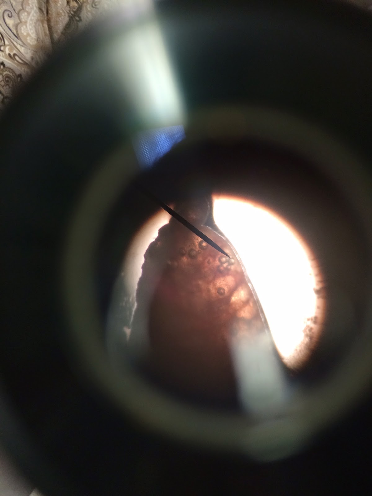
4) Select one lobe from the lungs to dissect. Using a teaser needle, make an incision down each side of the secondary bronchi. Do this in order to locate the tertiary bronchi, which branch off from the secondary bronchi.
5) Follow the tertiary bronchi with the teaser needle. You will be able to locate and observe the alveoli at the ends of the tertiary bronchi.
6) Once you have located the primary, secondary and tertiary bronchi, take a picture.
7) Now use the scalpel to remove a tiny piece of the end of the lobe you were just dissecting.
8) Take out the microscope and a glass slide. Place the piece of lung on the glass slide and place the glass slide under the microscope. Observe the alveoli under the microscope.
9) Take a picture of the microscopic view of the alveoli.
10) Place the heart, lungs and trachea back into the plastic bag.
Data and Observations: I found it quite easy to locate the primary, secondary and tertiary bronchi. It was very interesting to dissect a lung because I have always wondered what the inside of a lung looks like. I also enjoyed observing the alveoli under the microscope. At first I thought the air bubbles in the lung were the alveoli, but then my teacher corrected me; The white circles I saw were the alveoli and the black smaller circles were air bubbles. Looking at the lung under the microscope was very cool! All of Semara's (the cat's) organs seemed to be in very good shape, despite the fat around her heart. I definitely enjoyed this unit and cannot wait to continue dissecting! Pictures and data shown below.
*INCASE YOU CAN'T READ THE PICTURE, READ THE TEXT BELOW!
What did you learn from the Lung presentation that you did?
I learned the seriousness of cystic fibrosis. I learned that cystic fibrosis causes a defect in a gene that causes sticky, thick mucus to develop in tubes, ducts and passageways of the lungs, making it very difficult and painful to breathe. I also learned about the different chest physical therapy and machines that can help a person with cystic fibrosis breathe better, Most importantly I learned where to get treatment or a diagnosis if I or a loved one ever has symptoms of cystic fibrosis.


No comments:
Post a Comment